


8 Types of Dark Spots on Face: A Clinical Guide for 2026
Generic dark spot advice fails because it treats every brown mark as the same problem. It isn't. Hyperpigmentation is a visible endpoint, not a single diagnosis. The pigment may come from cumulative ultraviolet exposure, a hormonal signal, prior inflammation, friction, medication, or a lesion that shouldn't be self-treated at all.
Biologically, the common thread is melanin. Melanocytes increase pigment output when skin receives a trigger, and enzymes such as tyrosinase help drive that process. But the trigger matters more than is often appreciated. A flat sun spot, a patch of melasma, and a post-acne mark can all look similar in the mirror while responding very differently to acids, pigment inhibitors, lasers, or simple neglect.
That's why many “dark spot correctors” disappoint. They target the color you see, but not the mechanism creating it. If you want better outcomes, start with diagnosis, then match treatment to cause, depth, and skin reactivity. If you want a gentler product pathway, you can explore gentle, natural skin brightening, but the most significant impact comes from knowing which category of pigment you're dealing with.
The most useful way to understand the types of dark spots on face is as clinical case files. Each one has a pattern, a reason people fail to improve it, and a treatment logic that makes sense.
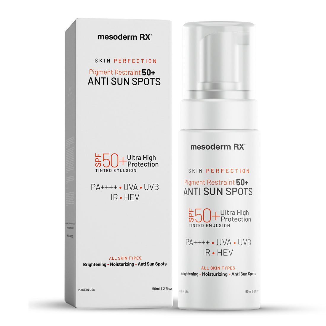
Featured Product
Pigment Restraint Ultra High Sun Protection
SHIELDS FROM UV DAMAGE AND SUN-INDUCED DARK SPOTS
Shop Now →1. Solar Lentigines (Sun Spots/Age Spots)
Solar lentigines are a record of repeated ultraviolet exposure, not a spot that appeared with age. They present as flat, sharply bordered tan to brown macules on chronically exposed areas, especially the cheeks, temples, forehead, and dorsal hands. A dermatology review cited by DERMASCOPE notes that solar lentigines are found in 90% of light-skinned people over age 60.
Case file logic matters here. Layer 1 is the mechanism. UV radiation drives localized melanocyte activity and increases melanin transfer into surrounding keratinocytes. Over time, that repeated signal creates stable pigment clusters that persist long after a tan would normally fade. In practice, these lesions often reflect both excess pigment production and epidermal changes caused by years of sun exposure.
Why treatment often stalls
The common failure pattern is simple. Patients target the visible mark and ignore the exposure pattern that keeps it active.
A single brightening serum rarely changes much if the person gets incidental UV on the drive to work, during walks, or near windows and never reapplies sunscreen outdoors. Another problem is product mismatch. Strong acids used too often can irritate the skin, while weak formulas used sporadically never generate enough turnover to fade established pigment.
Misidentification also slows progress. Freckles usually darken and lighten with seasons. Solar lentigines tend to stay visible year-round. If a lesion is raised, rough, asymmetric, or changing in color or border, it should not be managed as routine pigmentation until a clinician has looked at it.
Clinical clue: A flat, well-demarcated brown spot that remains in the same place through winter on a high-exposure facial area usually points to cumulative UV injury.
What works in practice
Layer 3 is treatment design. The goal is to suppress new pigment, remove pigment-bearing keratinocytes gradually, and stop daily UV from resetting the problem.
- Daily photoprotection: Broad-spectrum SPF 30 or higher is the baseline, and reapplication every 2 hours outdoors matters because ongoing UV exposure keeps melanocytes active.
- Measured resurfacing: AHA, BHA, or PHA used a few nights per week can improve epidermal turnover. The trade-off is irritation risk, especially in reactive skin, so frequency matters.
- Pigment control: Tyrosinase-inhibiting brighteners make more sense when paired with sunscreen than when used alone.
- Lesion review: Any spot with texture, bleeding, crusting, or visible change needs evaluation instead of more home treatment.
For patients building a pigment-focused UV strategy, Pigment Restraint Ultra High Sun Protection ($29.99, in stock) is described by the brand as a fast-absorbing, non-greasy formula designed to help reduce the look of visible sun damage while protecting against UVA, UVB, IR, and HEV-related light exposure.
For a broader treatment plan, Mesoderm RX also outlines how to get rid of dark spots with a structured routine. Exposure habits still decide whether results hold, especially around water, sand, and reflective surfaces, so practical sun safety tips for beachgoers are worth applying alongside skincare.
2. Melasma (Chloasma)
Melasma is a pattern diagnosis, not a single spot. It presents as broad, fairly symmetric brown to gray-brown patches, usually on the cheeks, forehead, nose, upper lip, or chin. The biology is different from an isolated sun spot because melanocytes are being pushed by more than UV alone. Hormonal signaling, visible light, heat, and baseline skin inflammation can all keep pigment production active.
That mechanism explains the relapse pattern clinicians see so often. Pigment may lighten for a few weeks, then return after pregnancy, a contraceptive change, outdoor heat exposure, or an irritating skincare cycle. The melanocyte was never fully quieted. The routine only thinned surface pigment for a short time.
A visual reference helps because melasma has a pattern distinct from isolated sun spots.

Why most melasma routines fail
Case file logic matters here.
Layer 1. Biological mechanism: Melasma often involves both epidermal and deeper dermal pigment, plus an overreactive melanocyte network. That is why it looks diffuse, why it can appear stubborn, and why high-friction treatment plans often backfire.
Layer 2. Common treatment failure: Patients often use stronger acids, more often, for longer than their skin can tolerate. The result is low-grade inflammation, barrier disruption, and more signaling for pigment production. Another common miss is treating melasma as a sunlight-only problem. Heat exposure, visible light, and hormonal drivers can keep patches active even when someone thinks they are being careful.
A realistic protocol controls pigment while limiting inflammation. That trade-off is the center of treatment. Melasma usually improves with steady suppression, not repeated cycles of irritation and recovery.
What a system-based melasma protocol looks like
Start with trigger control. Review heat exposure, hormonal shifts, and products that sting, peel, or leave the skin tight. Then build treatment in three lanes:
- Pigment suppression: Use ingredients that interrupt melanin formation rather than relying on exfoliation alone.
- Barrier-first resurfacing: Gentle keratolytics can help remove retained pigmented cells, but frequency has to match skin tolerance.
- Daily photoprotection: UV matters, but visible light and heat management also matter in melasma-prone skin.
For the resurfacing lane, AHA + BHA + PHA Skin Brightening Serum 30ml is described by the brand as containing kojic acid, glycolic acid, lactic acid, salicylic acid, lactobionic acid, hyaluronic acid, and hexapeptides, with a formulation intended to gently exfoliate and reduce the appearance of hyperpigmentation while improving tone and texture. Price and stock status were not provided in the source material, so they should be checked on the product page before purchase.
If melasma is the pattern on your face, use a plan written for that diagnosis rather than borrowing an acne or sun-spot routine. Mesoderm RX provides a more specific framework in its guide to melasma cream for face.
A short clinical explainer is useful here before you decide how aggressive to be:
3. Post-Inflammatory Hyperpigmentation (PIH)
PIH is the dark mark left after inflammation. Acne is the most familiar trigger, but eczema, psoriasis, irritation, friction, burns, and overly harsh procedures can do the same thing. The lesion heals. The pigment remains.
This category matters because the trigger is so common. In an AAD-linked acne context, acne affects up to 50 million Americans annually, which helps explain why post-inflammatory pigment is a major reason people keep searching for treatment after a breakout is already gone. Independent dermatology references also identify PIH as especially common in darker skin tones because melanocytes tend to react more strongly after inflammation.
What people get wrong
They attack PIH like active acne. That's the mistake. Once the spot is post-inflammatory, your first job is stopping the inflammation that created it. If breakouts are ongoing, if the cleanser burns, or if exfoliation is too frequent, you keep producing new marks while trying to fade old ones.
Another mistake is equating PIH with textural scarring. PIH is color. Scars are structural change. They can coexist, but they aren't the same problem.

A more effective order of operations
Treat PIH in this sequence:
- Stop the trigger: Control acne, dermatitis, friction, or product irritation first.
- Use low-irritation actives: Barrier damage will prolong the cycle.
- Add gradual resurfacing: Mild acid use can help lift residual pigment.
- Protect from UV: Sunlight darkens these marks and extends their life.
PIH improves fastest when skin is calm. Brightening inflamed skin is usually slower than calming it first.
If you're trying to set realistic expectations, Mesoderm RX has a practical guide on how long it takes for hyperpigmentation to fade. The key point is simple. Don't chase speed with stronger irritation.
4. Freckles (Ephelides)
Freckles are one of the easiest facial pigment patterns to misclassify. They're small, usually uniform, and often appear early in life rather than later. They darken with sun exposure and can soften when exposure drops.
That seasonal behavior separates them from many solar lentigines. If someone tells you the spots become much more obvious after a sunny period and lighten when they're less exposed, freckles move higher on the list.
Why the usual advice is incomplete
People often tell patients to “lighten freckles” without asking whether they should be treated at all. Freckles are benign pigment expression. The more relevant clinical question is whether the person wants less contrast, whether UV exposure is excessive, and whether there are any atypical lesions mixed in.
The treatment trade-off is also different. Because freckles are usually numerous and diffuse, spot-treating each one doesn't make much sense. Full-face management often works better.
What works and what doesn't
- Best approach: Strict daily sunscreen and sun-avoidance habits. Freckles respond strongly to exposure patterns.
- Reasonable support: Gentle brightening serums and measured exfoliation can reduce overall visual intensity.
- Poor strategy: Overcorrecting with harsh peels in someone prone to irritation.
A common real-world scenario is the fair-skinned patient whose nose and cheeks darken every summer despite using actives but not reapplying sunscreen outdoors. That's not a failed brightening product. It's an exposure-control failure.
5. Seborrheic Keratosis
Seborrheic keratosis is where “dark spot” self-diagnosis becomes risky. These lesions are usually benign, but they aren't simple pigment patches. They tend to be raised, waxy, stuck-on in appearance, and sometimes rough or scaly.
That texture changes the treatment logic completely. Topical pigment products won't flatten a three-dimensional lesion.

The diagnostic issue that matters
Consumer searches often lump every brown facial mark into hyperpigmentation. That's a problem because some lesions need professional evaluation rather than self-treatment. A dermatology overview notes the importance of screening lesions for warning signs such as asymmetry, irregular borders, color variation, diameter over 6 mm, and evolution over time.
If a “dark spot” is raised, newly changing, bleeding, itching in a new way, or visually atypical, the right next step is an exam, not an acid serum.
Clinical caution: Flat pigment can often be observed and treated conservatively. Raised lesions need a lower threshold for professional assessment.
What to do instead of experimenting
- Get it checked if it's new or changing
- Don't pick or scratch it
- Don't attempt home removal
- Use gentle skincare around it to reduce friction
A lot of wasted time in pigment care comes from treating non-pigment lesions as if they were melanin-only problems.
6. Erythema Dyschromicum Perstans (EDP/Ashy Dermatosis)
EDP is less common in general skincare conversations, which is why people often mislabel it as stubborn melasma, diffuse PIH, or “gray patches that won't brighten.” The color clue matters here. These patches often look gray-brown or ashy rather than just brown.
That shift in tone suggests you're no longer dealing with a routine sun-spot pattern. In practice, these cases usually need a more careful diagnostic approach because the exact cause can be less obvious, and progress tends to be slower.
Why common brightening plans disappoint
EDP often gets over-exfoliated. Someone sees persistent gray pigment, assumes the answer is stronger peeling, and then triggers more irritation. If inflammation is part of the process, that approach can keep the skin cycling without much visible gain.
Another issue is misclassification. If the diagnosis is wrong, the protocol will be wrong.
Practical treatment logic
A cautious plan usually makes more sense than an aggressive one:
- Confirm the diagnosis when possible
- Prioritize low-irritation brightening and antioxidant support
- Use sunscreen daily
- Avoid friction, heat, and unnecessary actives
A typical scenario is the patient with symmetric, ash-toned facial patches who has already tried multiple peels with little change and increasing sensitivity. That doesn't mean pigment is untreatable. It means the working diagnosis may need revision, and the skin may need calming before further correction.
7. Acanthosis Nigricans (Dark Neck/Velvety Hyperpigmentation)
Acanthosis nigricans is often discussed around the neck and body folds, but it belongs in this conversation because facial-border and fold-adjacent darkening can overlap with concerns people describe as “dark spots.” The key feature isn't just color. It's texture. The skin often looks velvety, thickened, and darker than the surrounding area.
That texture is the clue that this isn't ordinary superficial pigment alone. When I see a patient trying to bleach or scrub this kind of darkening off, I'm less interested in the brightener they chose and more interested in the underlying driver.
What people miss
This can be associated with underlying metabolic issues. If the pigmentation is velvety, concentrated in folds, and accompanied by thickening, skincare alone is incomplete care. The skin may improve somewhat with topical support, but the root issue needs medical review.
The right priority
- Get medical evaluation for possible systemic contributors
- Use gentle keratolytic and brightening support, not harsh scrubbing
- Reduce friction in affected areas
- Treat it as a texture-plus-pigment condition
This is one of the clearest examples of why the types of dark spots on face and nearby facial zones can't be sorted by color alone. Texture changes the diagnosis.
8. Perioral and Periocular Hyperpigmentation (Dark Circles and Mouth Shadows)
These are some of the most misread facial pigment complaints. “Dark circles” and “mouth shadows” are often treated as if they were a single pigment problem, but clinically they are a mixed-zone diagnosis. The color may come from epidermal melanin, dermal pigment, visible vasculature through thin skin, structural hollowing, chronic inflammation, or repeated friction. If the mechanism is wrong, the treatment plan fails.
That is why these areas fit a case-file approach better than a product-first approach.
Layer 1. Biological mechanism.
Periocular skin is thin, so vascular congestion and shadowing from the tear trough can read as pigment even when melanin is only a small part of the picture. Perioral darkening often has a different driver. Low-grade irritant dermatitis, lip licking, saliva exposure, toothpaste irritation, hair-removal trauma, and retinoid migration can keep melanocytes activated and the barrier unstable. In both zones, repeated inflammation increases pigment signaling. In darker skin tones, that inflammatory loop can produce persistent discoloration from relatively minor irritation.
Layer 2. Why treatment fails.
Failure usually starts with overcorrection. Strong acids, aggressive retinoids, and frequent rubbing damage the barrier in the exact areas that tolerate irritation poorly. Patients then interpret the extra darkness as “stubborn pigment” and intensify treatment again. Around the eyes, allergy-related rubbing is a common hidden driver. Around the mouth, dermatitis is often missed because the skin may look dry, shadowed, and brown rather than obviously red.
A broad clinical review from Harvard Health on hyperpigmentation causes, types, and treatments makes the larger point clearly. Hyperpigmentation management works better when trigger control, daily photoprotection, and carefully chosen topicals are used together. Over-exfoliation can worsen inflammation-driven pigment, especially in sensitive skin and skin of color.
Layer 3. System-based protocol.
- Identify the dominant signal first: pigment, vascular show-through, structural shadow, or irritation.
- Stabilize the barrier before escalating: reduce rubbing, pause irritating actives, and correct obvious contact triggers.
- Use lower-irritation brighteners: niacinamide, tranexamic acid, azelaic acid, arbutin, and carefully formulated vitamin C derivatives usually make more sense here than strong peel-style exfoliation.
- Introduce actives slowly: a few nights per week is often enough at the start.
- Treat anatomy accurately: hollowing and deep-set shadows do not respond like true hyperpigmentation.
For friction-linked darkening elsewhere on the body, Bikini Line & Inner Thigh Brightening Cream, Beach-Ready Skin in 30 Days is presented by Mesoderm RX as a dermatologist-tested vegan formula with tranexamic acid, kojic acid, arbutin, niacinamide, vitamin C, AHA, azelaic acid, and hexylresorcinol. Price and availability were not provided in the supplied catalog data, so they are not listed here. The clinical takeaway still applies to the face. Friction-driven darkening improves best when trigger reduction and pigment control are paired, rather than treating discoloration as a stain that responds to scrubbing alone.
9. Mixed Morphology Dark Spots
Some faces don't have one pigment type. They have several. A person may have lentigines on the temples, PIH on the cheeks, and broader background discoloration around them. Clinically, simplistic advice breaks down fastest in these situations.
A recent optical coherence tomography study classified dark spots into three structural categories based on rete ridge deformation at the dermal-epidermal junction, and after 2 months of full-face application in Asian women, the study measured a 7% reduction in melanin density inside dark spots and a 12% reduction in surrounding skin. That matters because spots that look similar on the surface may differ biologically.
Why this changes treatment decisions
If morphology differs, response differs. One spot may respond to resurfacing. Another may need melanin-signal suppression. A third may be too structurally distinct for over-the-counter care to do much.
Don't judge dark spots only by color. Distribution, border, depth, and texture often predict response better than shade alone.
What to do with mixed-pattern faces
Treat the face as a map, not a single lesion. Use full-face prevention, selective brightening, and conservative escalation. If you're documenting progress, consistent photos in the same lighting are more useful than memory.
Comparison of 8 Facial Dark Spot Types
| Condition | Implementation complexity 🔄 | Resource requirements ⚡ | Expected outcomes ⭐📊 | Ideal use cases 💡 | Key advantages ⭐ |
|---|---|---|---|---|---|
| Solar Lentigines (Sun Spots/Age Spots) | Low–Moderate: daily SPF + topical exfoliants/brighteners; consistency required | Moderate: SPF 50+, AHA/BHA/PHA serums; occasional in‑office procedures | Good: 50–75% fading in 8–12 weeks with strict UV protection | Sun‑exposed age spots in older individuals seeking non‑invasive correction | Responsive to topicals; highly preventable with sunscreen; benign |
| Melasma (Chloasma) | High: multi‑modal regimen + hormonal assessment and strict UVA protection | High: prescription depigmenters, professional procedures, long‑term maintenance | Moderate: 60–70% improvement possible; high relapse without ongoing care | Symmetric, hormone‑related facial patches (women of reproductive age) | Multiple therapy options; significant improvement when combined with hormonal management |
| Post‑Inflammatory Hyperpigmentation (PIH) | Moderate: gentle regimen plus trigger control and avoidance of irritation | Low–Moderate: mild exfoliants, antioxidants, SPF; some may need professional help | Good: 70–80% improvement in 3–6 months with proper care | Dark spots following acne, eczema, injury, especially in darker skin types | Preventable by avoiding trauma; responds well to gentle treatment; relatively faster recovery |
| Freckles (Ephelides) | Low: prevention‑focused with intermittent brightening | Low: daily SPF, gentle serums/exfoliants; minimal clinical intervention | Very good: 60–80% fading with strict sun avoidance and topicals | Hereditary, small sun‑reactive spots appearing from youth | Highly responsive to sun protection; easy maintenance; often cosmetically neutral |
| Seborrheic Keratosis | Low–Moderate: requires dermatologic evaluation; topical brighteners ineffective | Moderate–High: clinic procedures (cryotherapy, curettage, laser) for removal | Excellent (cosmetic) when removed: definitive clearance via procedures | Raised, waxy benign growths causing cosmetic concern or irritation | Definitive procedural removal available; benign and non‑spreading |
| Erythema Dyschromicum Perstans (EDP/Ashy Dermatosis) | High: prolonged multi‑modal therapy and specialist assessment | Moderate–High: ongoing topicals, antioxidants, strict SPF; possible specialist interventions | Moderate: 40–60% improvement over 6–12 months; some refractory cases | Chronic ash‑gray patches in darker skin types needing dermatologist care | Potential for gradual improvement with comprehensive brightening; generally stable once managed |
| Acanthosis Nigricans (Dark Neck/Velvety) | High: medical workup for metabolic causes + topical texture management | High: systemic treatment (metabolic screening, weight/glucose management) plus specialized topicals | Good: 60–80% improvement when underlying metabolic factors treated over months | Velvety fold hyperpigmentation linked to insulin resistance/obesity | Reversible when root cause addressed; systemic benefit beyond skin |
| Perioral & Periocular Hyperpigmentation (Dark Circles/Mouth Shadows) | Moderate–High: sensitive‑area protocols; gradual escalation to avoid irritation | Moderate: specialized gentle serums, SPF, adjunctive vascular/supportive treatments | Good: 50–70% melanin improvement; vascular component often 30–50% | Delicate periocular/perioral pigmentation with high cosmetic impact | Visible overall appearance improvement; tailored gentle regimens available |
Your Protocol for Pigment Clarity: From Diagnosis to Results
Results come from matching treatment to mechanism. This step is often overlooked. They buy a “dark spot serum,” use it on every brown mark they can see, and then conclude that pigment care doesn't work. The failure was diagnosis.
Start by identifying the lesion pattern. Flat, well-defined marks on high-exposure areas often point toward cumulative UV damage. Broad, symmetric patches suggest melasma. Residual spots after acne or irritation usually fit PIH. Raised, waxy, or changing lesions move out of the self-treatment category quickly.
Then deal with the trigger. Sun-driven pigment requires rigorous daily sunscreen and reapplication when you're outdoors. Inflammation-driven pigment requires stopping the breakout, rash, friction, or product irritation that keeps provoking melanocytes. Hormonal pigment requires realistic expectations because a good topical routine can help, but it can't erase an ongoing trigger by itself.
From there, build a system. In practical terms, that usually means combining carefully chosen exfoliants, pigment-regulating actives, and barrier-respecting support rather than relying on one harsh formula. Sensitive skin does better with steady, tolerable use than with aggressive cycling. Darker skin tones often need even more caution because inflammation itself can deepen discoloration.
One market reality is worth noting because it reflects what patients already do. Grand View Research reported that the age spots segment held the largest revenue share at 36.6% in 2024, while energy-based therapy held 68.4% of treatment share. That tells you two things. First, sun-related pigment remains a dominant concern. Second, many people still need an at-home maintenance plan even when procedures are part of care.
The best protocol is the one you can sustain without irritating your skin. Daily photoprotection, disciplined use of actives, and realistic timelines outperform frantic product switching every time. If you want a consumer-friendly overview of product-based fading, this guide on how to fade dark spots with corrector cream can be a useful companion to a more diagnosis-driven routine.
Mesoderm RX is one relevant option for people who want hydroquinone-free, multi-active routines built around brightening, resurfacing, and sun protection. The main point remains the same regardless of brand. Identify the spot correctly, remove the trigger when possible, and use a structured routine long enough to judge it fairly.
If you want a routine built around targeted actives, daily UV defense, and practical pigment correction for sensitive or uneven skin, explore Mesoderm RX.